Thursday, 3 August 2017
Health problems

- Eye infections
- Pug dog encephalitis
- Inflammation of the brain
- Inflammation of the cornea
- Ulcers on the cornea
- Dystocia
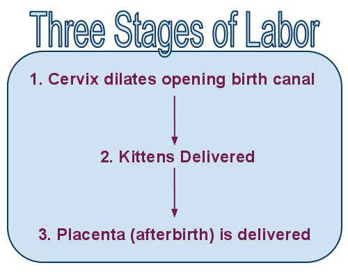
Labour stages:
The gestation period for both dogs and cats is for approximately for 60 days. A key sign of parturition is a sudden drop in body temperature by about 1 degree Celsius The body temperature however returns back to normal or increases a little at the time of parturition. Dogs under stress or first time reproducers can delay the delivery of their pups by a day without causing any harm to them. When this happens, there is a temperature drop for the entire period until parturition begins. Labour in cats and dogs can be divided into 3 main stages.
First stage labour:
The first phase of parturition can last in cats and dogs between 6-12 hours. At this time cervical dilation and vaginal constriction takes place, but there is no signs of uterine or abdominal contractions are observed. During this phase, a behavioral change is observed and the bitch is often found to get restless, nesting, panting and licking her vagina now and then.
Second stage labour:
The second stage of labour last between 3 to 12 hours. It can sometimes even extend to 24 hours depending on the litter size. The average litter size for most dogs and cats is between 5 to 8 neonates. The time period between each neonate varies between 15 minutes to 6 hours, though one hour is more common as well as normal. Once a fetus is born, it should take anywhere between 5 minutes to 2 hours before the next is delivered. A time duration of more than 4 hours is risky and must be addressed. All pups and kittens are born with the amnion (a membrane) around them. Furthermore, most of the pups and kittens are born in cranial presentation with a few born in caudal presentation.
Third stage of labour:
The third stage of labour involves the removal/ expulsion of the placenta. Every pup/kitten is delivered with a placenta. However 2 or more pups born followed by the expulsion of a few placenta is also normal(canine placenta separation). During this phase a darkish green colored fluid is also seen to ooze out. This fluid is called uteroverdin and it originated from the junction between the placenta and the endometrium of the uterus. This fluid is only seen to be expelled during or post delivery, not prior.
Monday, 26 March 2012
Diagnosis of dystocia
The diagnosis of dystocia ultimately relies on the subjective assessment of labour progression. Continuous contractions of the myometrium should expel a neonate every half an hour, with a maximum duration stretched to 4 hours if there are weak contractions. However the more the delivery duration is delayed the greater the risk for the neonate and the more stress the dam is in.
A clinical exam should primarily include a minimum database of blood test in order to rule out systemic illness like hypoglycemia and hypocalcemia. Uterine rupture will also present as a problem and hence a physical examination of the vulva and the caudal vagina can be done. Uterine rupture will usually present as a physical shock and possible toxemia- pale gums, rapid heart rate, weak pulses and abnormal blood work. If the blood work show signs of hypoglycemia or hypocalcemia then this problem must be addressed immediately. However, if the blood work is normal then it might be uterine inertia and oxytocin ca be administered.
Radiography is very useful as it will determine the number of neonates in the uterus as well as their position. Ultrasonography is also particularly beneficial as it can detect individual foetal heart rates.
Oxytocin
A very important thing to remember is that oxytocin does not increase the strength of uterine contractions, but instead the frequency. The effect of oxytocin lasts for approximately 30-90 minutes, and the second dose can be administered after 30 to 60 minutes. However, it must be noted that 30% of the bitches do not deliver the neonate even with the administration of oxytocin. Hence in this case surgical intervention via caesarean is warranted.
Preparation of arrival:
· Clean bedding in a quiet location is ideal along with a few seats for the owners to sit as they usually like to present when their pet is being monitored.
· Plenty of clean towels
· Water based lubricant
· Skin prep and tape and appropriately sized catheters
· Oxytocin and needles and syringes
· Ultrasound: if available, must be set up and ready to go
· X-Ray: the X-Ray must be set up according to the size of the abdomen of the patient and ready to go
· Monitoring equipment: in order to monitor the pulse rate, ECG, BP etc.
· oxytocin, needles and syringes
· Surgical equipment: 1.anesthetic machines, ET tubes and drugs
2. plenty of towels for neonate resuscitation
3. extra hemostats for umbilicus
4. plenty of warm lavaging saline
· Humidcrib/oxygen induction chamber or warm location for the neonates, post delivery.
Considerations of anesthesia for caesareans:
1. During parturition, the uterus usually exerts extra pressure on the lungs and diaphragm, hence decreasing the tidal volume, thereby decreasing anesthetic requirements and increasing the respiratory rate.
2. The patient’s head should never be inclined downwards during delivery as this will greatly increase the pressure on the diaphragm which will result in an increase in abdominal pressure on the diaphragm and this can greatly compromise ventilation.
3. Pregnant females also have delayed gastric emptying-they tend to regurgitate and possibly aspirate more than normal patients.
4. The surgeon must be ready soon after the patient is anesthetized, as exposing neonates to depressive drugs can be dangerous and decrease their chances of survival.
5. Hypotension: clipping the patient prior GA, can reduce the overall time of GA.
Subscribe to:
Posts (Atom)